
160 X Ray Anatomy Positions Ideas Anatomy X Ray Radiology Technologist

160 X Ray Anatomy Positions Ideas Anatomy X Ray Radiology Technologist

General Anatomy And Radiographic Positioning Terminology Radiology Key

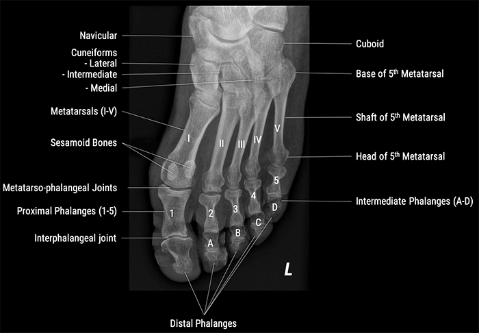
Foot X Rays

160 X Ray Anatomy Positions Ideas Anatomy X Ray Radiology Technologist

160 X Ray Anatomy Positions Ideas Anatomy X Ray Radiology Technologist
The choices we have include the posterior-anterior x-ray or the PA x-ray the lateral chest x-ray 000030 the anterior-posterior or AP x-ray the decubitus view the lordotic view the inspiration expiration series chest fluoroscopy dual-energy chest x-ray digital tomography or tomosynthesis.
Names of x ray views. You need to stay still when you are having an x-ray. The following are common types of x-rays. The preferred examination is the posterior-anterior x-ray.
IMV in-house Radiographer Kat Evans commented At IMV we understand that capturing a great X-ray isnt just about having the best equipment. An X-ray tube is a simple vacuum tube that contains a cathode which directs a stream of electrons into a vacuum and an anode which collects the electrons and is made of tungsten to evacuate the heat generated by the collision. 152 PELVIS X-Ray Pelvis 153 X-Ray Hip 154 X-Ray Sacroiliac Joint 155 UPPER EXTREMITY UPPER EXTREMITY X-Ray Shoulder 156.
Anteroposterior known as AP lateral skyline. An X-ray generator generally contains an X-ray tube to produce the X-rays. Possibly radioisotopes can also be used to generate X-rays.
Occlusal x-rays are most commonly used by pediatric dentists to check on the growth and formation of the teeth and jaw bone. BSM Orthopaedic Standard X-Ray Views 1. The easy to follow guides aim to help practices by providing a quick easy-to-use reference guide to getting the best radiographic views possible every time.
Ideally a shoulder radiograph series will provide adequate views of the clavicle acromioclavicular joint ACJ glenohumeral joint GHJ and the scapula. Lying on one side. The x-ray machine takes the image from beneath the chin for a view of the lower teeth and jaw or from above near the nose for the upper teeth and jaw.
X-Ray Chest 148 X-Ray Apical Lordotic View 149 X-Ray Sternum 150 X-Ray Ribs Oblique 151 ABDOMEN X-Ray KUB. PROCEDURE DESCRIPTION CPT CODE Chest 1 View 71010 Chest 2 Views 71020 Chest Minimum 4 Views 71030 Chest Special Views 71035 Ribs Unilateral 2 Views 71100 Ribs Unilateral 2 Views with PA CXR 71101 Ribs Bilateral 3 Views 71110 Sternum Minimum 2 Views 71120. The beam is aimed from dorsoproximal to palmarodistal at a 65 degree angle to.

Chest X Ray Positioning

Pin On Anatomy And Physiology

Foot Series Radiology Reference Article Radiopaedia Org

