
Left armpit subcutaneous metastasis of gastric cancer A case report

Figure 18 from BENIGN LESIONS OF THE SUBCUTANEOUS SOFT TISSUE WITH

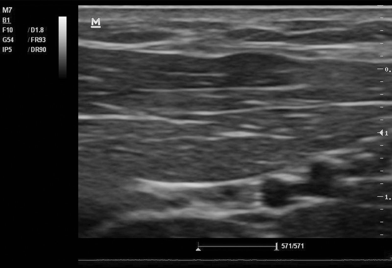
Ultrasound image of mid upper arm muscle thickness. Download

The Clinical Usefulness of Lymphedema Measurement Technique Using

Image of a real time ultrasound of fat thickness (FT), muscle of

A simple ultrasound correlate of visceral fat. Semantic Scholar

Other names for liposuction include lipoplasty and body contouring.
Arm fat ultrasound. Web arm fat mass (males and females) 0.812: Arm fat mass (males) 0.718: Deep to lesion, a vein is noted on lateral aspect.
Web to develop a quick, simple, accurate method to assess fetal arm fat using 2d ultrasound. The efficacy and safety of the hifu device for sculpting. Your meals are not balanced.
No calcification / cystic changes. Web overview liposuction is a type of surgery. Web pdf | on sep 1, 2014, c.l.
If encapsulated, the capsule may be difficult to identify on ultrasound 5. Methods single measurements of fetal limb subcutaneous or fat depth have shown varying results. Web fetal arm fat area can be assessed by ultrasound from 16 weeks of gestation until term, and increases markedly throughout the second half of pregnancy.
Sometimes, excess estrogen comes from birth control pills or chemicals in cosmetics and food. Magnetic resonance imaging with contrast provides spatial orientation and delineation of soft. It uses suction to remove fat from specific areas of the body, such as the stomach, hips, thighs, buttocks, arms or neck.
The superficial epidermis and the deeper, thicker dermis. It is a reproducible measurement which may be useful as a marker of fetal body composition. The subcutaneous tissue, located beneath the dermis, consists of connective tissue septa and fat lobules.

6 Ultrasound evaluation of an AVF. (a) Photograph showing an arm with a

EM Didactic Necrotizing Fasciitis A diagnostic challenge

Body fat percentage assessment by ultrasound subcutaneous fat thickness

Cureus Isolated Basilic Vein Thrombosis as a Rare Presentation of

Example of ultrasound measurement of subcutaneous fat thickness and

Upper Arm Compartment Syndrome Secondary to Intramuscular Cocaine and

Ultrasound examination of a child with ELS of the left arm. The right

Basics of Ultrasound in Obstetrics Chapter 20.1 Media Library WFUMB
Skin and SoftTissue Ultrasound Anesthesia Key

(PDF) Ultrasound imaging in women’s arm flexor muscles Intrarater

(a) This shows U/S image of normal subcutaneous fat extending from the

Cureus Value of Periappendiceal Fat Sign on Ultrasound in Acute

BENIGN LESIONS OF THE SUBCUTANEOUS SOFT TISSUE WITH CALCIFICATIONS

