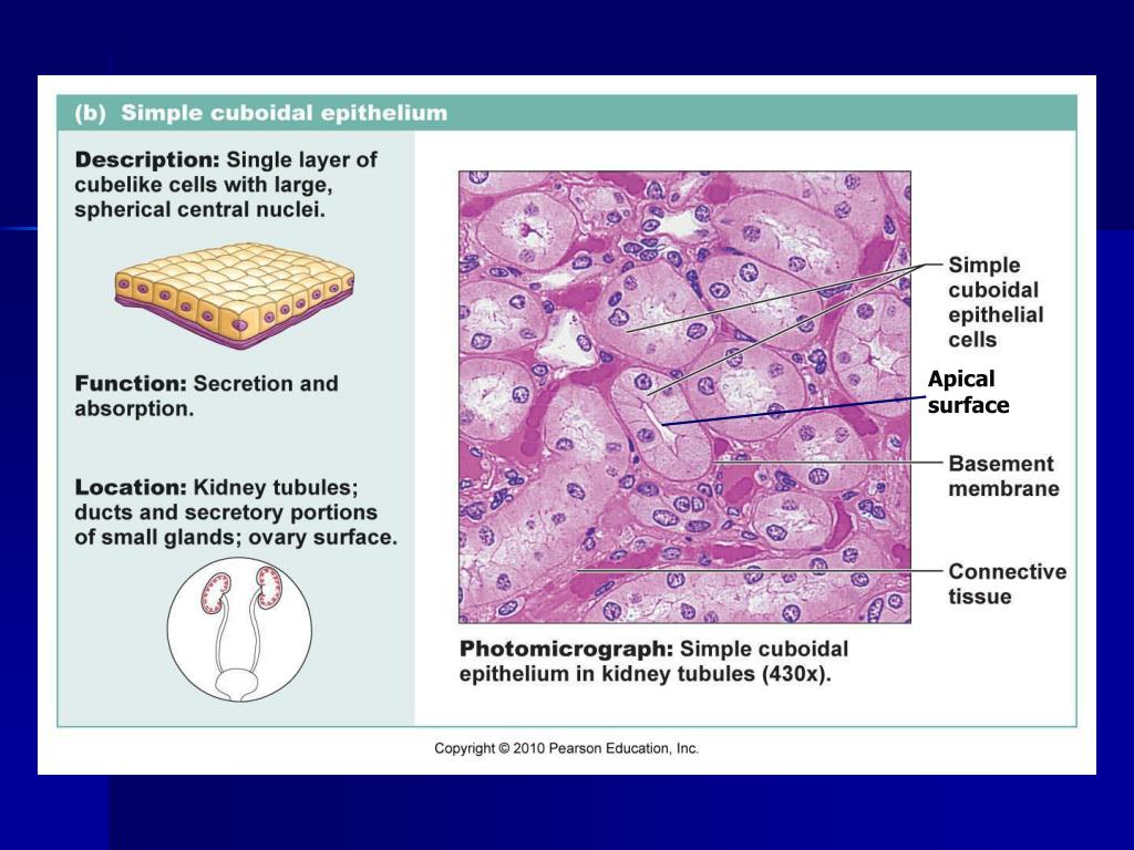
The surface of an epithelial cell that faces the lumen.
Apical surface of a cell. Airway surface liquid (ast), primarily composed of mucus gel and water, surrounds the airway lumen with a thickness thought to vary from 5 to 10 mm. The apical surface is closest to the base of the cell; Apical surface apical surface definition.
Of, at, or being the apex | meaning, pronunciation, translations and examples The apical surface is closest to or on the luminal surface. Cell lineages derived from the apical cell will progressively differentiate to form cotyledons, shoot meristem, hypocotyl axis, and embryonic root (figure 4).by contrast, the basal cell will divide.
The distal or apical boundaries of. 12 words view all related. Epithelial tissue contains a layer of.
The apical surface is sometimes covered with cilia. Epithelia cells are polarized with an apical surface that faces the lumen of a tube or the external environment and a basal surface that attaches to the basement membrane. The apical surface is the upper side of the epithelial cell that faces the external.
Homeostasis of many epithelial tissues involves the addition of new cells from basally positioned progenitors. Examine the process by which a new cell integrates into an. The intestinal cells are joined at the.
Apical surface in oxford dictionary of biochemistry and molecular biology (2) length: Ast lies on the apical surface of.


