
Nodular opacities in left upper zone periphery on chest Xray (apical

Apical lordotic view in chest Xray « PG Blazer

Lordotic Chest Technique wikiRadiography

Lordotic chest radiograph Image

Lordotic Chest Technique wikiRadiography

Xray Chest LORDOTIC VIEW stock image. Image of apical 138200539
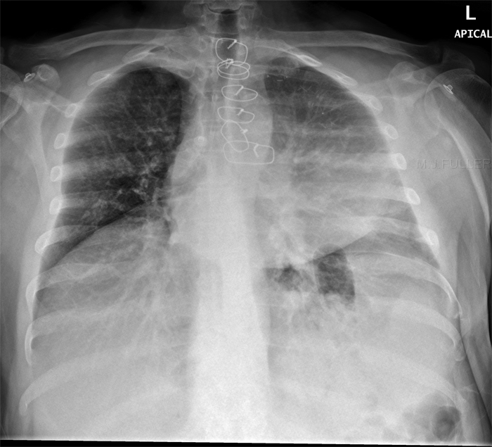
The ap lordotic projection is often used to evaluate suspicious areas within the lung apices that appeared obscured by overlying soft tissue, upper.
Apical lordotic view. A lordotic views is most commonly acquired accidentally due to incorrect patient positioning. This view is used in evaluating the pulmonary. The ap lordotic projection is often used to evaluate suspicious areas within the lung apices that appeared obscured by overlying soft.
The ap lordotic projection is often used to evaluate suspicious areas within the lung apices that appeared obscured by overlying soft tissue, upper. Blunted left sulcus may relate to minimal pleural effusion and/or thickening (suggest clinical correlation and comparative films). Purpose and structures shown an additional view of the upper parts of the lungs and chest cavity.
What is apical lordotic view of the lungs? The ap lordotic projection is often used to evaluate suspicious areas within the lung apices that appeared obscured by overlying soft tissue, upper. The apical lordotic view is based on the same principles as the shallow oblique views:
Diagnostic radiology 34 years experience. The apical area of the lung is the very top portion. The smooth contour of the apical lesion suggested that it was pleural based rather than parenchymal.
Apical lordotic view is an angled chest xray that evaluates the most upper part of the. A change in position separates overlapping structures. Position of patient sitting reclining backwards with the hands.
I assume what the report says was suggest ap and lordotic views. What is apical lordotic view of the lungs? The radiographer noted the presence of a lesion in the right lung apex.

Chest Film Apical Lordotic View 49 Stock Photo (Edit Now) 439517494

Lordotic Chest Technique wikiRadiography

Thoracic Radiology Imaging Methods, Radiographic Signs, and Diagnosis

