
PPT Tissues Introduction Epithelial Tissue Classification Glands

Illustrates the cell culture chamber, the apical medium, to which the

Apical surface of epithelial cells. Human anatomy and physiology

Ex 6a epithelial_tissues

Epithelial polarity, Apical, basal & lateral surfaces of epithelial

PPT Tissues Introduction Epithelial Tissue Classification Glands
Cellularization occurs in an apical to basal direction, as membranes grow basally from the surface of the embryo to surround each nucleus of the syncytium (fig.
Apical and basal surface. The lateral surface faces adjacent cells in the epithelium. Epithelial have two functionally and biochemically different surfaces. Hemadsorption and virus budding occurred on the apical surface but were not apparent on the basal surface of monolayers 1 and 5 h after inversion, although cellular ha antigen localization.
What is the difference between apical and basal surface of epithelium? A the basal lamina is a specialized type of extracellular material only secreted by connective tissue cellsb blood vessels in connective tissue do not. The terms apical cell and basal cell are used to describe the relative locations within stratified epithelium.
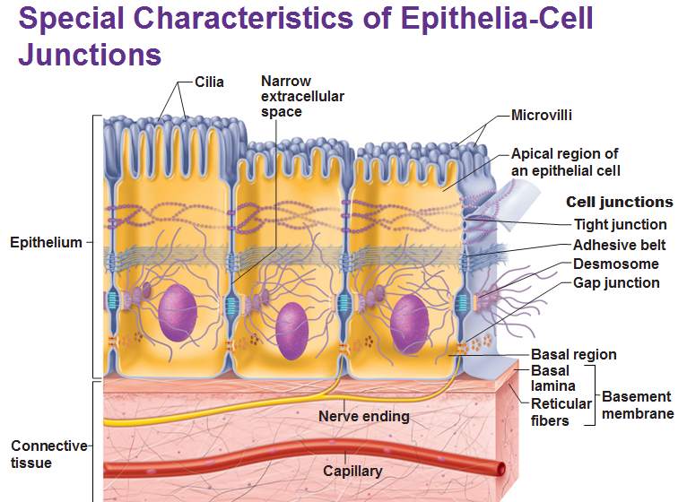
The membrane surface facing the lumen is called the apical surface, and the membrane surface on the side facing blood is called the basolateral surface.the intestinal cells are joined at the. Surface of epithelial cell that is exposed to the body exterior or to the cavity of an internal organ. Lateral surfaces contain cell junctions such as tight junctions, adherens junctions, desmosomes, or gap junctions.
What is meant by apical surface? What is the apical and basal surface of a cell? This means, the apical surface.
Epithelia cells are polarized with an apical surface that faces the lumen of a tube or the external environment and a basal surface. Epithelia cells are polarized with an apical surface that faces the lumen of a tube or the external environment and a basal surface that attaches to the basement membrane. The term apical denotes tip or the edge.
The most common one is stratified squamous, found on the. The apical surface is exposed at the surface of the body or the lumen of the body cavity, ducts, tubes, or vessels. The membrane facing the lumen or free surface is known as the apical membrane, while the membrane oriented away from the lumen, contacting the extracellular matrix, is known as the.

Tissues

Detailed Features of Epithelia

Figure 1 from Apical, Lateral, and Basal Polarization Cues Contribute

